


Content warning: mention of autopsy and graphic images of human anatomy
One cool opportunity I didn’t know about before coming to Princeton was the Princeternship program. Princeternships are a unique chance to get insight into a career of interest through one or more Princeton alumni. They take place over winter break and can range from a few days of shadowing to a few weeks of working on a project. This past year, more than 180 in-person and virtual Princeternships were available in the fields of Arts, Culture, Media & Entertainment; Engineering, Science & Technology; Business; Healthcare; Social Impact; and Law. I had the privilege of participating in a two-day virtual Princeternship at the Children’s Hospital of the King’s Daughters, hosted by Dr. Alice Werner. Together with four other Princeterns, I learned about Dr. Werner’s specialty, pediatric pathology, and got a glimpse into the operation of the hospital’s numerous departments.
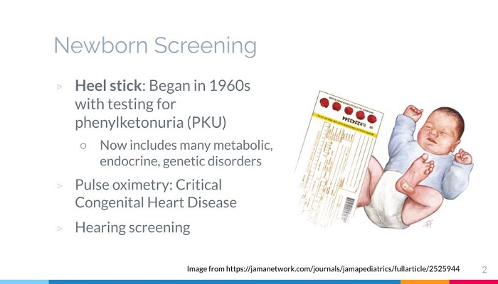
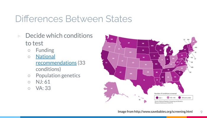
Prior to the Princeternship, Dr. Werner assigned each of us a topic to prepare a 10-15 minute presentation on, and we each gave our presentation when it was relevant to what we were discussing. I presented on the heel stick, a type of newborn screening, and the differences in screening in different states.
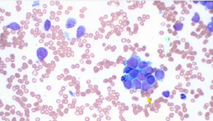
We were first introduced to some of the equipment and analytical techniques used in the hospital’s medical laboratory. One of my fellow Princeterns presented on the MALDI Biotyper, which identifies microorganisms using mass spectrometry. We spent the majority of our time observing slides of all kinds of samples under the microscope, including blood, cerebrospinal fluid and tumor biopsies. The virtual format was convenient because I could join from the comfort of my home and did not need to arrange travel or accommodation for during and after the Princeternship. Since I would have had to leave home several weeks earlier if the Princeternship were in-person, I was also able to enjoy some more time with my family. In addition, since Dr. Werner does a lot of work on the microscope and the computer, it was easy to see what she was looking at, as opposed to having six people crowded around one computer screen.
On the first day, Dr. Werner “brought us to work” and took us through a normal morning as a clinical pathologist. She brought up the medical history and lab results of the patient whose sample she was tasked with analyzing, then showed us the slide under the microscope and explained what she saw. For example, I learned that reactive lymphocytes in the blood, identified by their large size and turquoise cytoplasm, are an indication of viral infection. We sometimes went back to the doctor’s note or lab results to piece together an explanation of what we were seeing. It was fascinating to participate in Dr. Werner’s thought process in real time, and I was impressed by all the types of body fluids we can observe under the microscope and the variety of clues they can give about one’s health and the potential cause of their disease. It felt like going behind the scenes of a normal doctor’s visit: literally, because Dr. Werner entered notes which the doctor interacting with the patient would use to develop their treatment plan, and metaphorically, because we could dive right into the cells at the root of the problem.
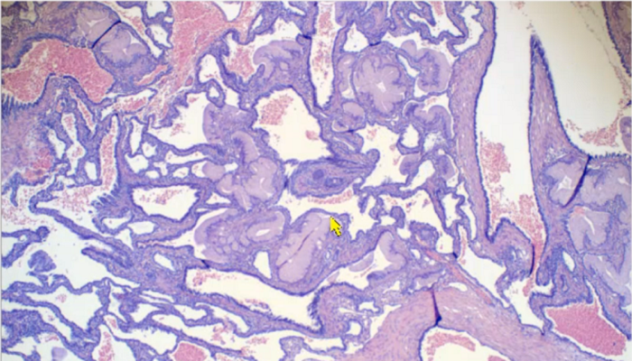
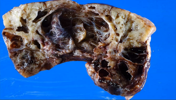
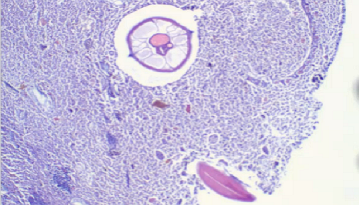
On the second day, we attended the hospital’s Daily Safety Briefing where representatives from each department reported any concerns from the last 24 hours. For the remainder of the day, Dr. Werner showed us anatomical pathology slides she had been collecting as well as the corresponding MRI and ultrasound scans to give us more context to the cases. As an anatomical pathologist, Dr. Werner sometimes performs autopsies and she showed us an image of a deceased newborn baby with gastroschisis, a defect where some of the baby’s intestines are not enclosed in the body. She then showed us the autopsy picture exposing the baby’s internal organs, and I must admit, I was both shocked and mesmerized. Part of me was desperate to look away because it felt wrong to see a baby like this, but another part of me still wanted to learn what Dr. Werner had to share. As a pre-medicine student, I will likely have to face this conflict often in the future. I hope to always hold a deep respect for the patient and their family and gratitude for their willingness to allow us to learn everything we can from their loss. I believe that image will stick with me for years to come because I was reminded of the fragility of life which I usually take for granted.
This Princeternship taught me about a specialty that I knew almost nothing about and gave me an appreciation for medical labs and the technicians, chemists, engineers and pathologists who make it possible for us to analyze specimens and thereby understand illnesses much better. Although I realized that I am probably not interested in pursuing pathology because I would like to interact with patients and do a bit more with my hands, I learned that pathology is a fascinating and extremely important specialty which also allows providers to have a regular work schedule.
A huge thank you to the Center for Career Development for this enlightening opportunity, and I hope to participate in another Princeternship in the future!