In a previous post, I described my geological field work during June and July in Namibia. I helped a graduate student in the Department of Geosciences, Akshay Mehra, with his research, which focuses on studying the first bio-mineralizing organisms about half a billion years ago. These organisms are especially interesting because they immediately precede the earliest known animals, so they could be the direct ancestors of the first animals. Nobody really knows much about them, though, so our field work focused mainly on collecting samples of rock containing the fossilized shells of one particularly abundant species, Cloudina. We eventually collected almost 1,600 pounds of samples, which miraculously made it to Princeton. Now, with field work done, we're about to start actually getting a glimpse of these tiny, tubular creatures that lived in those half-billion year-old reefs.

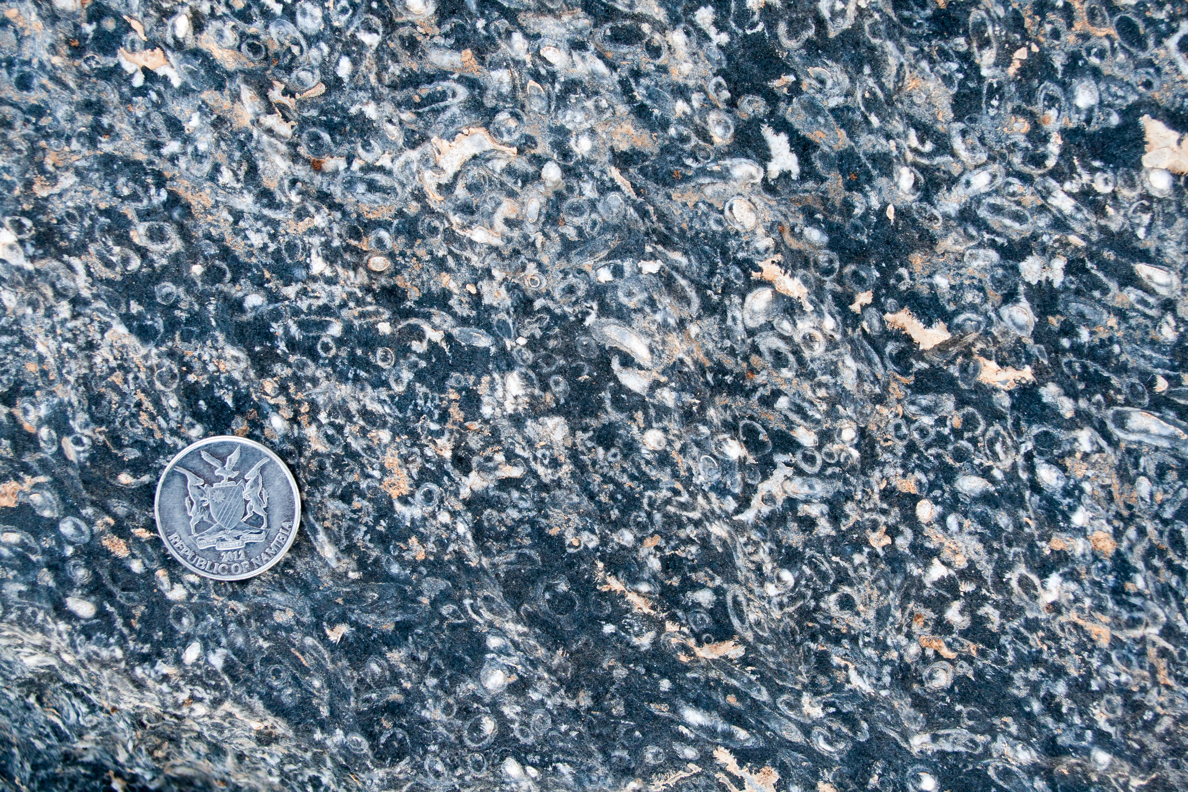
But how much can you learn about a 540 million-year-old, tube-shaped organism when all you can see in the surface of a rock are cross-sections through its often poorly preserved shell? Other types of fossils are easy to extract, but in the case of Cloudina, the fossils are actually the same exact material as the rock surrounding them; there's no way to dissolve them out or anything like that. All that actually makes the fossil recognizable from the rock around it is a slight color difference resulting from mild chemical change that only affected the shells.

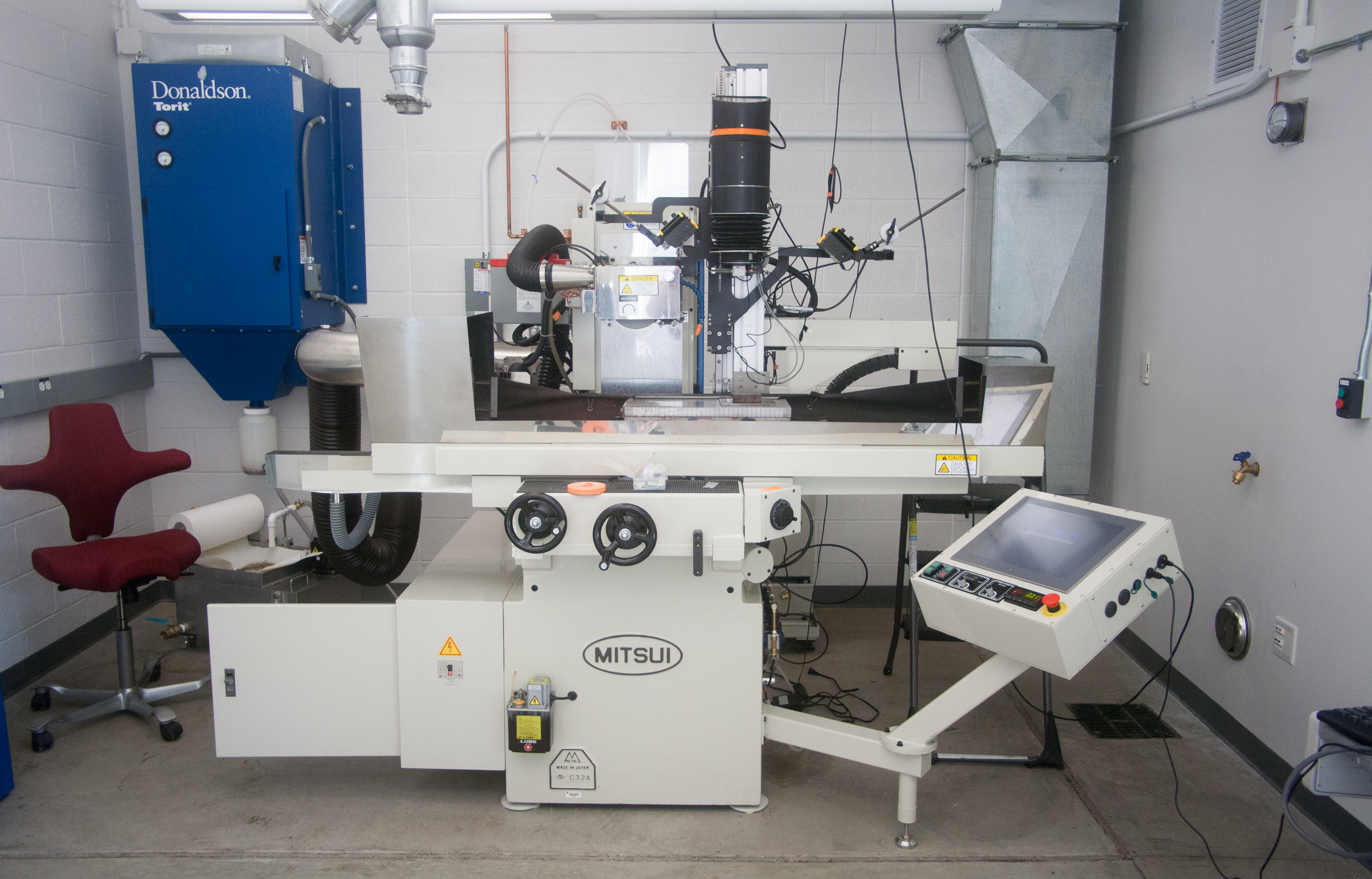
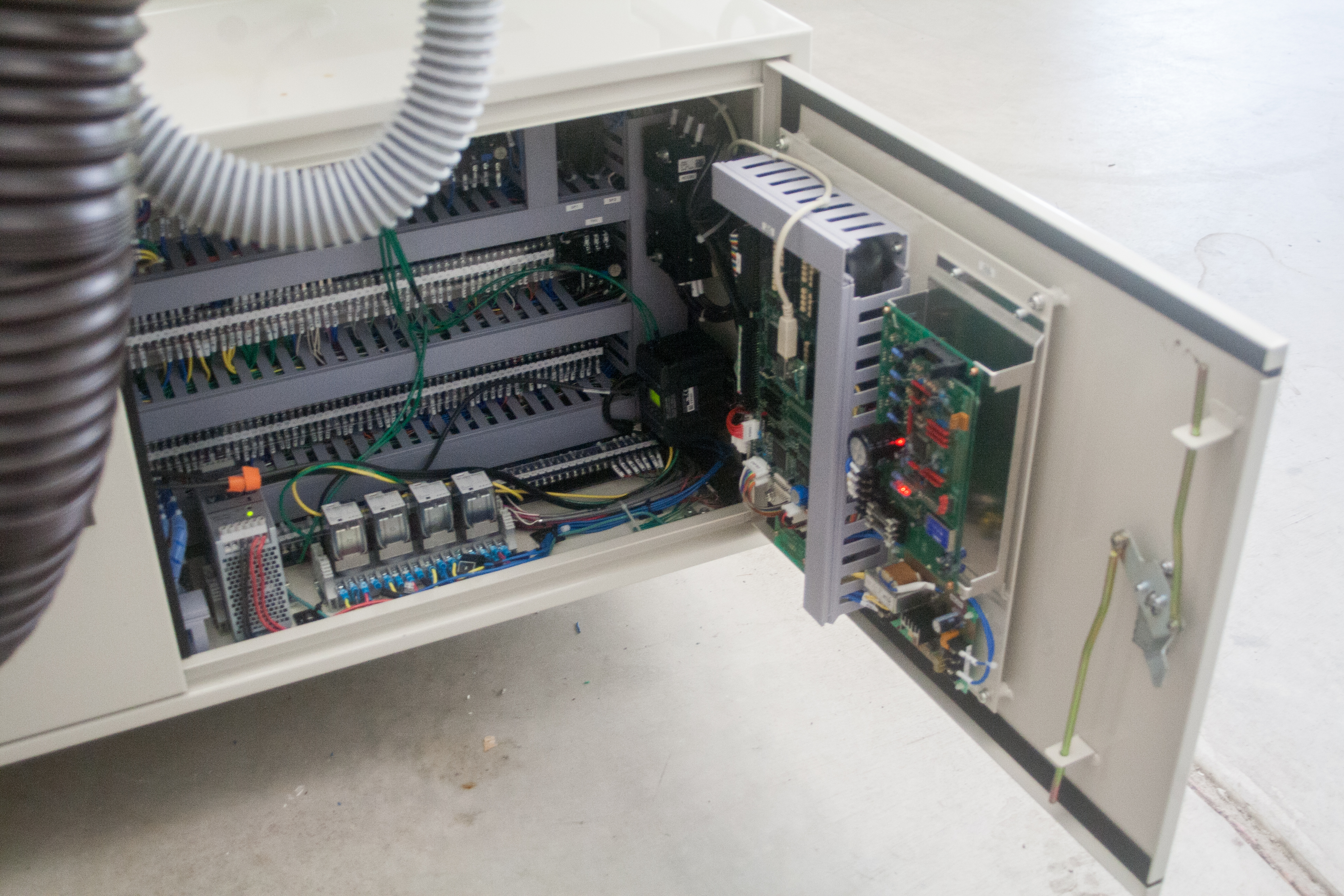
This piece of equipment is basically a machining grinder with several additions, including an 80 megapixel imaging sensor and controllers that interface it with a computer. The basic idea is that you take troublesome samples, like the ones we collected, and grind tiny layers off the sample. With each layer, you take an insanely high resolution photo of the surface, then grind again, then take another picture, and so on until the entire sample has been ground to dust. Yes, the sample is totally and literally pulverized, but what you have instead is hundreds of cross-sections through it, which you can then connect in three dimensions to create a 3D model of the sample. The magic of this technique is that you can see how the fossils look on the inside of the rock, and with a little bit of computer learning, you could even train the computer to recognize individual fossils and compute statistics about fossil density and orientation.
[embed]http://vimeo.com/72207631[/embed]

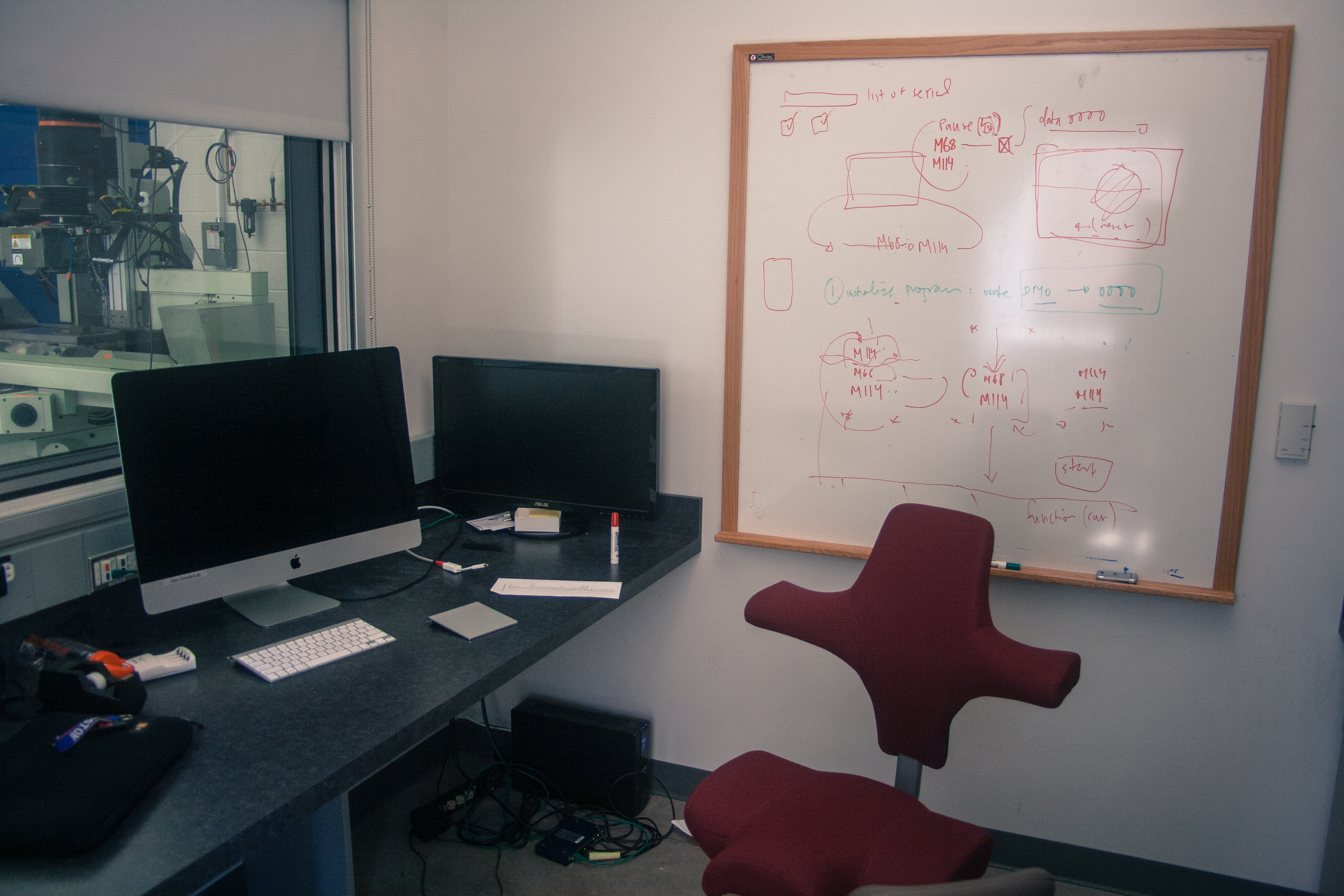
Honestly, just a year ago, I would never have guessed that I'd be looking at fossils of some of the earliest life-forms. Even beyond that, my work in the lab has taught me skills I've always wanted to know: how to interface different types of electric equipment with serial communication, all-purpose coding, and image analysis, to name a few. In just a few days we're going to attempt our first fully automated grind, which has never been done before, and it makes me proud to know I've contributed to something new in the scientific world.


